In recent years, particular interest has developed in molecular biology applied to the field of dermatopathology, with a focus on nevi of the Spitz spectrum. From 2014 onwards, an increasing number of papers have been published to classify, stratify, and correctly frame molecular
[...] Read more.
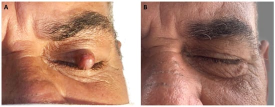
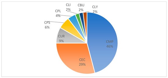
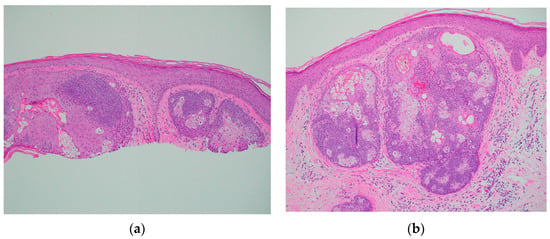
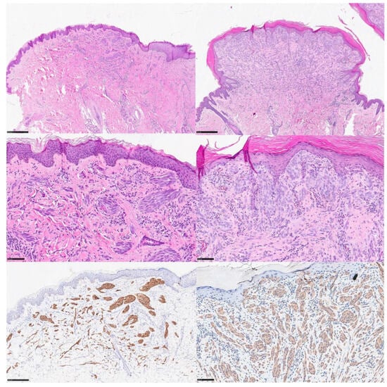
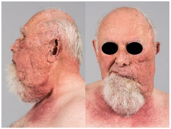
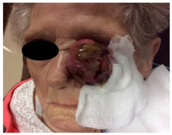
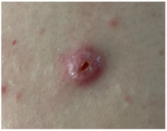
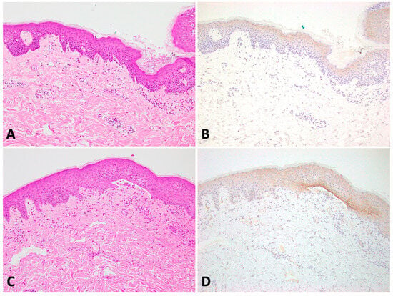
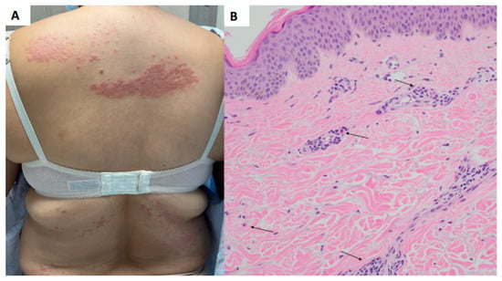
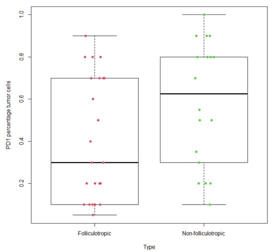
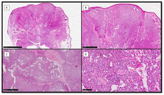
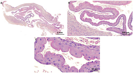
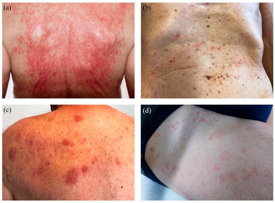
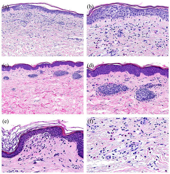
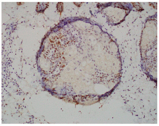
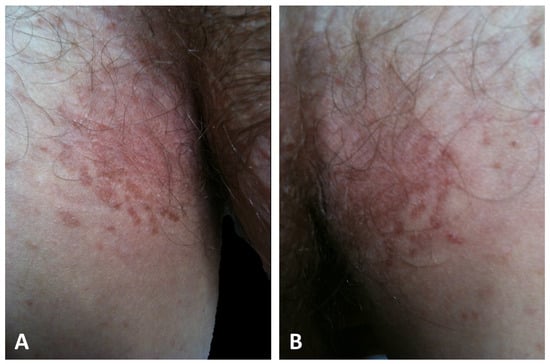
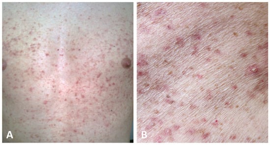
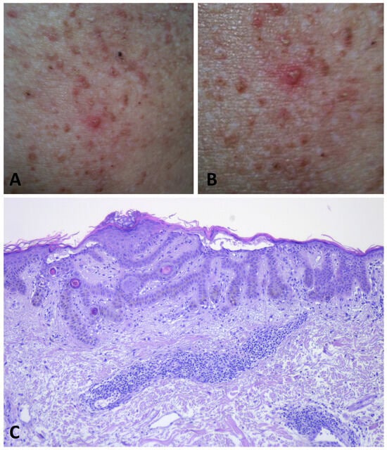
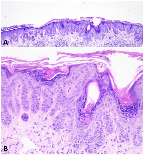
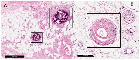
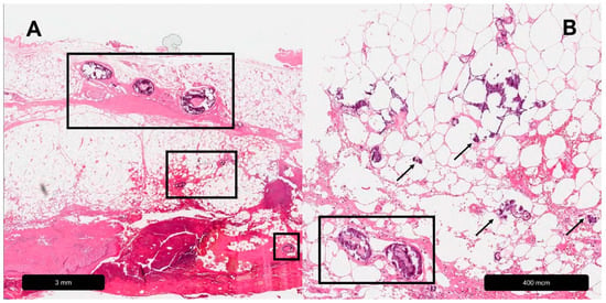
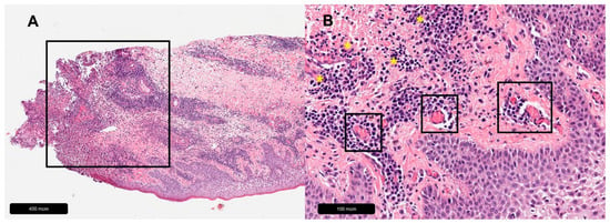
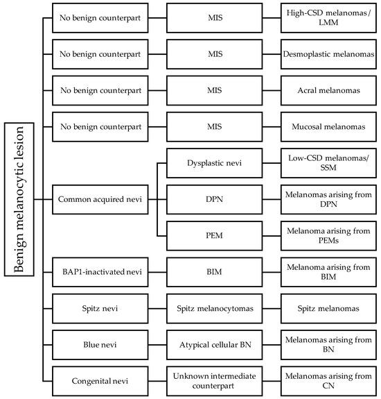
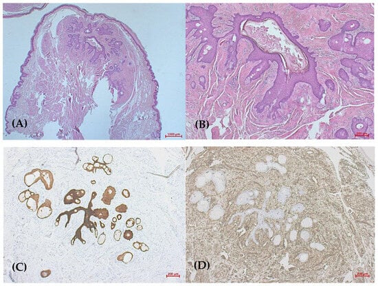
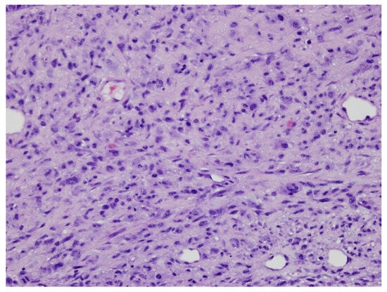
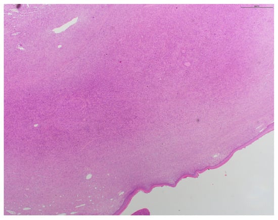
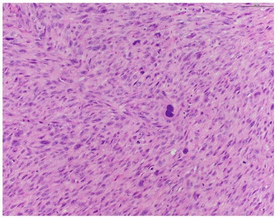
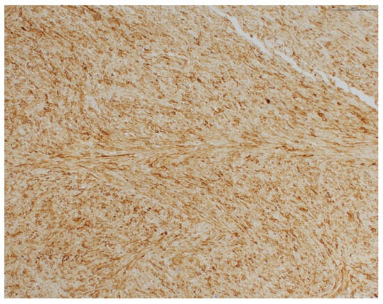
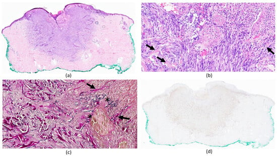
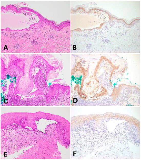
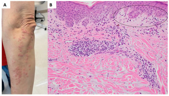
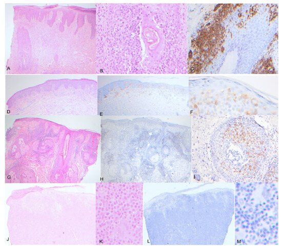
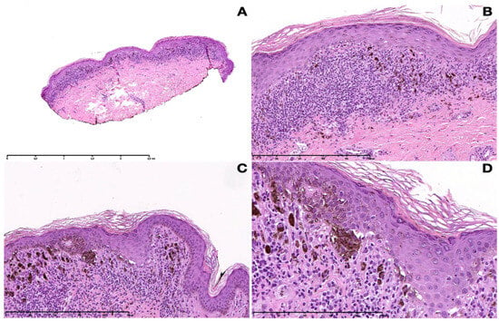
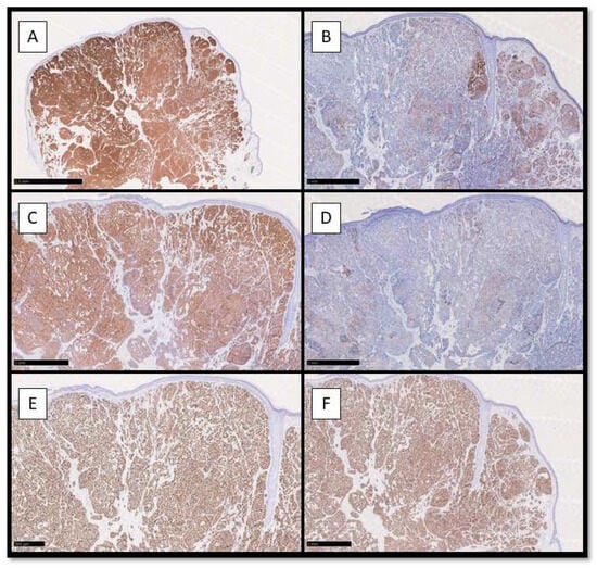
In recent years, particular interest has developed in molecular biology applied to the field of dermatopathology, with a focus on nevi of the Spitz spectrum. From 2014 onwards, an increasing number of papers have been published to classify, stratify, and correctly frame molecular alterations, including kinase fusions. In this paper, we try to synthesize the knowledge gained in this area so far. In December 2023, we searched Medline and Scopus for case reports and case series, narrative and systematic reviews, meta-analyses, observational studies—either longitudinal or historical, case series, and case reports published in English in the last 15 years using the keywords spitzoid neoplasms, kinase fusions, ALK, ROS1, NTRK (1-2-3), MET, RET, MAP3K8, and RAF1. ALK-rearranged Spitz tumors and ROS-1-rearranged tumors are among the most studied and characterized entities in the literature, in an attempt (although not always successful) to correlate histopathological features with the probable molecular driver alteration. NTRK-, RET-, and MET-rearranged Spitz tumors present another studied and characterized entity, with several rearrangements described but as of yet incomplete information about their prognostic significance. Furthermore, although rarer, rearrangements of serine–threonine kinases such as BRAF, RAF1, and MAP3K8 have also been described, but more cases with more detailed information about possible histopathological alterations, mechanisms of etiopathogenesis, and also prognosis are needed. The knowledge of molecular drivers is of great interest in the field of melanocytic diagnostics, and it is important to consider that in addition to immunohistochemistry, molecular techniques such as FISH, PCR, and/or NGS are essential to confirm and classify the different patterns of mutation. Future studies with large case series and molecular sequencing techniques are needed to allow for a more complete and comprehensive understanding of the role of fusion kinases in the spitzoid tumor family.
Full article


